Immunofixation Electrophoresis (IFE) Test: M-Proteins, High/Low Results, Symptoms & Complete Guide
Overview
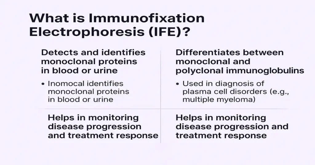
Immunofixation Electrophoresis (IFE), also known as Immunoelectrophoresis (IEP), is a specialized laboratory test used to detect and precisely identify abnormal antibodies, especially monoclonal proteins (also called M-proteins), in blood or urine. These proteins are produced when a single group of plasma cells behaves abnormally and starts making one identical antibody in excess.
IFE is most commonly associated with plasma cell–related conditions such as multiple myeloma, Waldenström’s macroglobulinemia, MGUS, primary amyloidosis, and light-chain disorders. From a laboratory perspective, this test is considered highly specific because it not only confirms the presence of an abnormal protein but also clearly identifies its exact type—for example IgG, IgA, IgM, or light chains (kappa or lambda).
In routine practice, IFE is usually performed after a Serum Protein Electrophoresis (SPEP) test shows a suspicious band or spike. IFE then acts as the confirmation step, helping doctors understand what that abnormal protein actually represents.
Where Abnormal Proteins Are Produced in the Body
The abnormal proteins detected on IFE originate from plasma cells, which are antibody-producing cells derived from B-lymphocytes and located mainly in the bone marrow.
Under normal conditions, plasma cells produce a wide variety of antibodies (polyclonal antibodies) to fight different infections. This diversity is a sign of a healthy immune system.
In certain disorders, however, a single plasma cell clone becomes dominant. That clone multiplies and produces only one type of antibody in large amounts. This single-type antibody is what we call a monoclonal protein or M-protein.
Different diseases tend to produce different patterns. For example, excess IgG or IgA is commonly seen in multiple myeloma, IgM in Waldenström’s macroglobulinemia, and free light chains in amyloidosis or light-chain disease. IFE helps the laboratory clearly identify which pattern is present.
Main Functions and Importance of the IFE Test
Detection of monoclonal proteins
The primary role of IFE is to confirm whether a monoclonal protein is present. This distinction is important because monoclonal patterns are often linked with plasma cell or lymphoid disorders rather than routine infections.
Differentiation between monoclonal and polyclonal patterns
From a diagnostic viewpoint, this is critical. Polyclonal patterns usually reflect inflammation or infection, while monoclonal patterns raise suspicion for plasma cell diseases. IFE provides clarity when routine tests are inconclusive.
Support in diagnosing plasma cell and lymphoid disorders
IFE is a key diagnostic tool in conditions such as multiple myeloma, MGUS, Waldenström’s macroglobulinemia, amyloidosis, and light-chain diseases. It does not stand alone but fits into a broader diagnostic framework.
Monitoring disease course
In patients already diagnosed, IFE helps track whether the abnormal protein persists, disappears, or changes over time. Clinically, this supports assessment of disease stability or progression.
Complements other investigations
IFE works best when interpreted alongside SPEP, UPEP, serum free light chain assays, and, when needed, bone marrow studies. Together, these tests give a reliable picture of plasma cell activity.
Causes of Low / Negative IFE Results
A negative IFE result means no monoclonal protein was detected, which is generally reassuring.
This can occur in healthy individuals with a normal immune response. It may also be seen when abnormal proteins are present at levels too low to be detected, such as in very early or mild disease states. In many cases, symptoms prompting testing turn out to be related to conditions unrelated to plasma cell disorders.
From a laboratory standpoint, a negative IFE suggests there is no current evidence of a monoclonal antibody pattern.
Symptoms of a Negative Result
A negative IFE result itself does not cause symptoms. Any symptoms experienced by the patient usually arise from other medical conditions rather than from a plasma cell disorder.
Causes of High / Positive IFE Results
A positive IFE result confirms the presence of a monoclonal protein, indicating abnormal plasma cell activity.
This pattern is commonly seen in multiple myeloma, where excessive IgG, IgA, or light chains are produced. Waldenström’s macroglobulinemia typically shows an IgM monoclonal pattern. MGUS presents with a smaller amount of monoclonal protein and is often stable but monitored over time.
Primary amyloidosis is associated with free light chains that deposit in tissues. In some cases, chronic immune stimulation or autoimmune conditions may produce unusual antibody patterns that require careful interpretation.
Symptoms of High / Positive IFE Levels
Symptoms are related to the underlying condition rather than the test result itself.
In plasma cell disorders, patients may experience bone pain, fatigue, recurrent infections, anemia, or kidney-related issues. Disorders involving IgM may present with circulation or nerve-related symptoms. Amyloid-related conditions may show organ-specific signs such as swelling, weakness, or heart and kidney involvement.
From a clinical perspective, IFE helps explain why these symptoms may be occurring, rather than causing them.
Reference Ranges
IFE does not use numerical reference ranges. Results are interpreted based on visual band patterns.
A normal result shows broad, diffuse polyclonal immunoglobulin patterns. An abnormal result shows a sharp, well-defined band in a specific immunoglobulin or light-chain lane, such as IgG, IgA, IgM, kappa, or lambda.
Quantification of protein levels is done using other tests like SPEP or serum free light chain assays.
Sample Type
IFE can be performed on serum (blood) or urine samples.
Serum testing is most common for detecting intact immunoglobulins. Urine testing, usually via a 24-hour collection, is particularly useful for detecting free light chains, also known as Bence Jones proteins. Proper sample collection is important for accurate interpretation.
Test Preparation
IFE testing generally requires minimal preparation. Fasting is not needed, and regular medications are usually continued unless otherwise advised.
Patients should inform their doctor about kidney disease, chronic infections, or autoimmune symptoms. For urine testing, careful adherence to 24-hour collection instructions improves reliability.
When to Consult a Doctor
Medical evaluation is important if there are unexplained symptoms such as persistent bone pain, frequent infections, ongoing fatigue, weight loss, anemia, kidney abnormalities, or nerve-related complaints.
IFE is also recommended when routine blood tests show high total protein, raised globulin levels, or abnormal SPEP findings. These results often prompt further assessment to rule out or confirm plasma cell disorders.
Important Word Explanations
- Paraprotein – Another name for a monoclonal protein.
- Monoclonal protein (M-protein) – An abnormal antibody produced by a single plasma cell clone.
- Electrophoresis – A laboratory technique that separates proteins based on size and charge.
- Immunofixation – A method that uses antibodies to identify specific immunoglobulin types.
- Plasma cells – White blood cells responsible for antibody production.
- Bence Jones proteins – Free light chains found in urine, often associated with myeloma.
~END~
Related Posts
None found