Overview
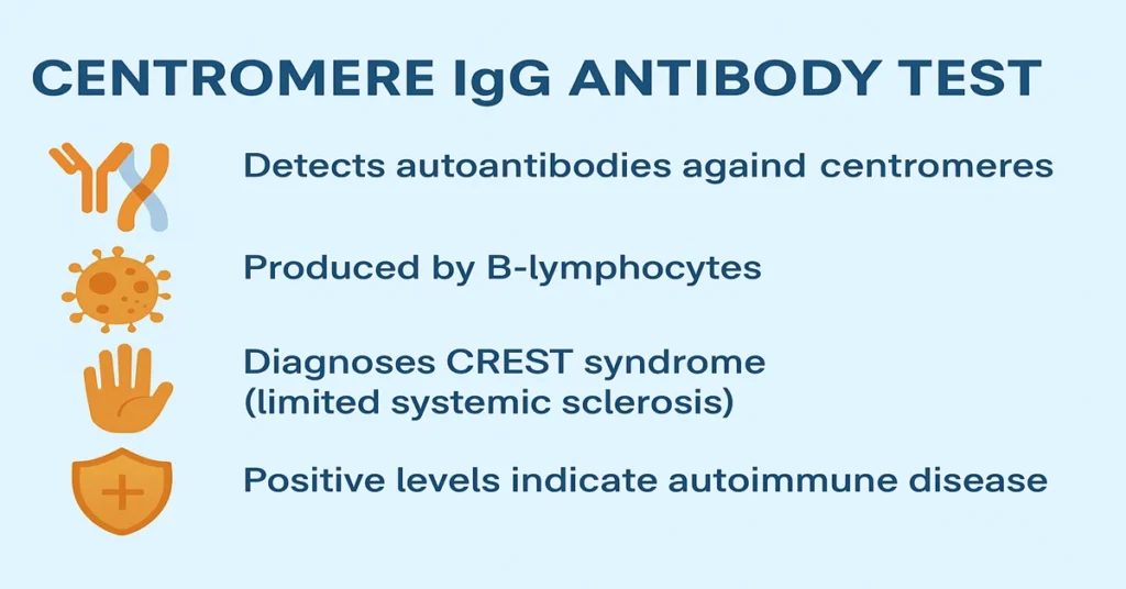
The Centromere IgG Antibody Test is a specialized blood test that detects autoantibodies directed against centromere proteins, which are located in the central region of chromosomes. These antibodies are most strongly associated with limited cutaneous systemic sclerosis, commonly referred to as CREST syndrome, and are occasionally seen in certain other autoimmune or connective tissue disorders.
From a clinical perspective, this test helps doctors recognize and classify autoimmune diseases at an early stage. When interpreted alongside symptoms and other laboratory findings, it supports accurate diagnosis and long-term disease planning.
What Is the Centromere IgG Antibody Test?
This test measures IgG antibodies that react against centromere proteins. Centromeres are essential structures that ensure chromosomes separate correctly during cell division and maintain genetic stability.
In autoimmune conditions, the immune system mistakenly identifies these normal cellular structures as foreign. As a result, antibodies against centromere proteins are produced. Their presence signals an autoimmune process rather than an infection.
Clinically, this test is most often used in the evaluation of:
- Limited cutaneous systemic sclerosis (CREST syndrome)
- Occasionally, primary biliary cholangitis (PBC)
- Rare overlap or connective tissue autoimmune conditions
Where Are Centromere Antibodies Produced in the Body?
Centromere antibodies are produced by B-lymphocytes, also known as plasma cells. These cells normally produce antibodies that protect the body from infections.
In autoimmune diseases, immune regulation is disrupted. The body loses tolerance to its own tissues, leading to the formation of autoantibodies. In this case, antibodies are formed against centromere proteins, which are otherwise normal components of human chromosomes.
Main Functions and Importance of the Test
Centromere proteins themselves play a vital role in normal cell division, but antibodies against them do not serve any protective function. Instead, they act as markers of autoimmune activity.
The Centromere IgG Antibody Test is clinically important because it:
- Supports diagnosis of CREST syndrome, a slower-progressing and more limited form of systemic sclerosis
- Helps differentiate limited from diffuse systemic sclerosis, which have different patterns of organ involvement and prognosis
- Provides diagnostic clarity when symptoms such as Raynaud’s phenomenon, skin tightening, or esophageal problems are present
From a laboratory standpoint, the presence of these antibodies often points toward a specific disease pattern rather than generalized autoimmune activity.
Causes of Low or Negative Levels
A low or negative result means centromere antibodies were not detected. This usually indicates:
- No autoimmune response directed against centromere proteins
- CREST syndrome is unlikely
However, a negative result does not completely exclude autoimmune disease. Other connective tissue disorders may involve different antibodies that are not detected by this test.
Symptoms of Low or Negative Levels
A negative result does not cause symptoms on its own. It is simply a laboratory finding.
If symptoms of autoimmune disease persist despite a negative result, doctors often investigate further with additional antibody tests, such as ANA, anti-Scl-70, or anti-RNP antibodies, to look for alternative autoimmune causes.
Causes of High or Positive Levels
A positive Centromere IgG antibody result indicates autoimmune activity targeting centromere proteins. The most common clinical associations include:
- Limited cutaneous systemic sclerosis (CREST syndrome), where these antibodies are frequently present
- Primary biliary cholangitis (PBC), in a subset of patients
- Other connective tissue overlap syndromes, less commonly
In clinical practice, a positive result is interpreted carefully in combination with symptoms and other immune markers.
Symptoms of High or Positive Levels
Symptoms are related to the underlying autoimmune condition rather than the antibody itself. Common clinical features may include:
Skin and soft tissue findings
- Tight or thickened skin, especially on fingers and face
- Shiny or stiff skin over the hands
- Calcium deposits under the skin (calcinosis)
Vascular symptoms
- Raynaud’s phenomenon, with color changes in fingers or toes triggered by cold or stress
Digestive involvement
- Difficulty swallowing
- Chronic acid reflux due to esophageal motility issues
Other features
- Telangiectasia (small visible blood vessels on the skin)
- Fatigue or itching in patients with associated liver disease
Reference Ranges
Reference ranges vary slightly between laboratories, but commonly reported values include:
- Negative: less than 20 AU/mL
- Borderline / Equivocal: 20–25 AU/mL
- Positive: greater than 25 AU/mL
Borderline results are often reassessed or confirmed with repeat testing or alternative methods.
Sample Type and Test Method
Sample type:
- Blood (serum)
Testing methods:
- ELISA (Enzyme-Linked Immunosorbent Assay)
- Indirect Immunofluorescence (IFA)
These methods reliably detect and quantify centromere antibodies. Fasting is generally not required for this test.
Test Preparation
No special preparation is needed.
Patients should inform their doctor about ongoing symptoms, known autoimmune conditions, or family history of similar diseases. Medications are usually continued as prescribed unless specifically advised otherwise.
When to Consult a Doctor
Medical evaluation is recommended if you experience:
- Persistent Raynaud’s phenomenon
- Progressive tightening or thickening of the skin
- Difficulty swallowing or long-standing reflux symptoms
- Unexplained fatigue, joint stiffness, or skin changes
- Visible small red blood vessels on the skin
Early recognition of conditions such as CREST syndrome allows for better monitoring and prevention of long-term complications.
Important Word Explanations
- Autoantibodies: Antibodies that mistakenly target the body’s own tissues
- Centromere: The central part of a chromosome that ensures correct separation during cell division
- Systemic sclerosis: An autoimmune condition causing skin and organ thickening
- CREST syndrome: A limited form of systemic sclerosis with characteristic clinical features
- Raynaud’s phenomenon: Color changes in fingers or toes due to blood vessel spasm
- ELISA: A laboratory method used to detect and measure antibodies in blood
~END~
Related Posts
None found